
Figure 3.
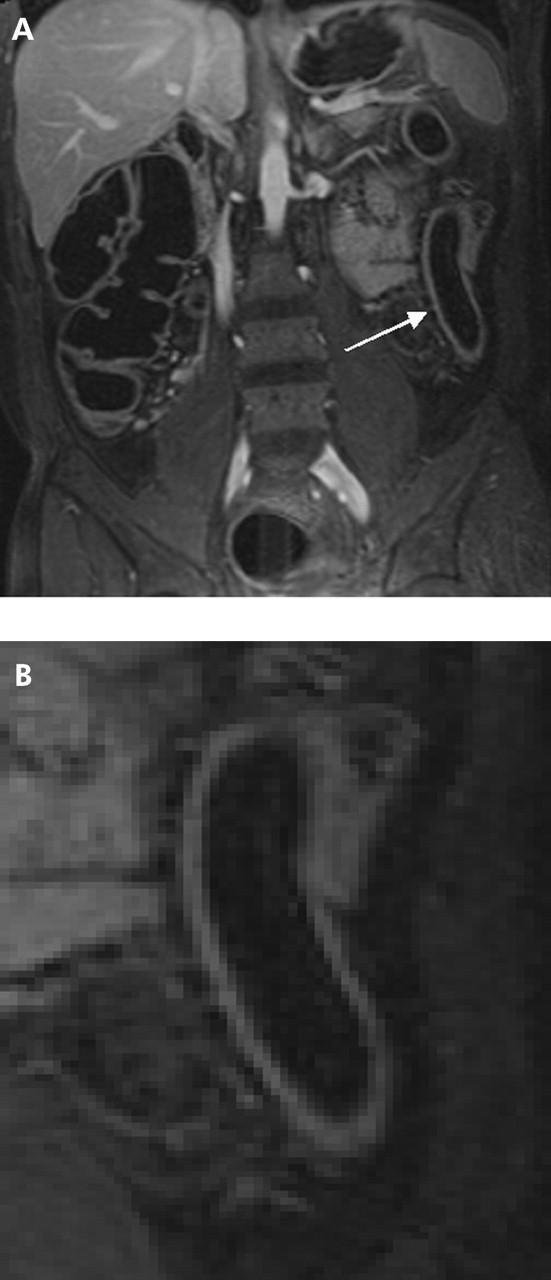
(A) T1 weighted three dimensional GRE image (TR/TE 3.1/1.1) of a 47 year old male patient with known ulcerative colitis. Magnetic resonance colonography diagnosed an inflammation in the descending colon (arrow). (B) Detailed display of the descending colon of the same patient which displays loss of haustral markings and slight bowel wall thickening. Due to the absence of lymph nodes as well as normal contrast uptake in the colonic wall, inflammation was rated as slight. This was confirmed by subsequent endoscopy.