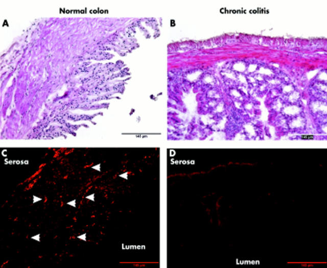
Figure 4.
Sympathetic innervation of the colon in normal (A, C) and colitic (B, D) mice. (A, B) Haematoxylin-eosin staining of colonic tissue. (C, D) In subsequent cross sections, sympathetic nerve fibres were stained by antibodies against tyrosine hydroxylase and secondary fluorescence labelled antibodies. White arrowheads indicate bundles of sympathetic nerve fibres in normal colon. All micrographs were taken at 100× magnification.

