
Figure 6.
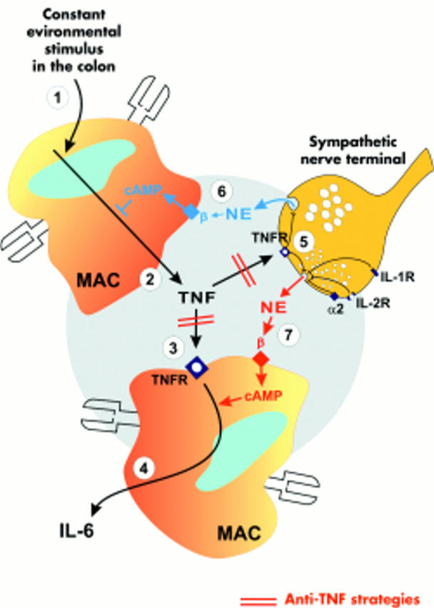
Model of colonic interleukin 6 (IL-6) regulation by tumour necrosis factor (TNF) and norepinephrine (NE) in normal colon. A line with an arrow (a bar) indicates a stimulatory (inhibitory) pathway. Constant environmental stimuli provoke local TNF secretion from, for example, macrophages (MAC (1)). (2) TNF is secreted into the local microenvironment and binds to TNF receptors (TNFR) on macrophages (3) and sympathetic nerve terminals (5). TNF stimulates downstream IL-6 secretion which becomes the readout parameter of TNF regulation (4). Under conditions without anti-TNF, the blue β-adrenoceptor (βAR) pathway (6) dominates the red βAR pathway (7), which leads to NE induced inhibition of TNF and subsequently IL-6 secretion. In the presence of anti-TNF, the influence of TNF is completely blocked and the effects mediated by the blue βAR pathway are negligible. Under these conditions, the red βAR pathway dominates and IL-6 is stimulated via an increase in extracellular NE and intracellular cAMP. Other cytokines such as IL-1 or IL-2 inhibit NE release from sympathetic nerve terminals.