
Figure 4.
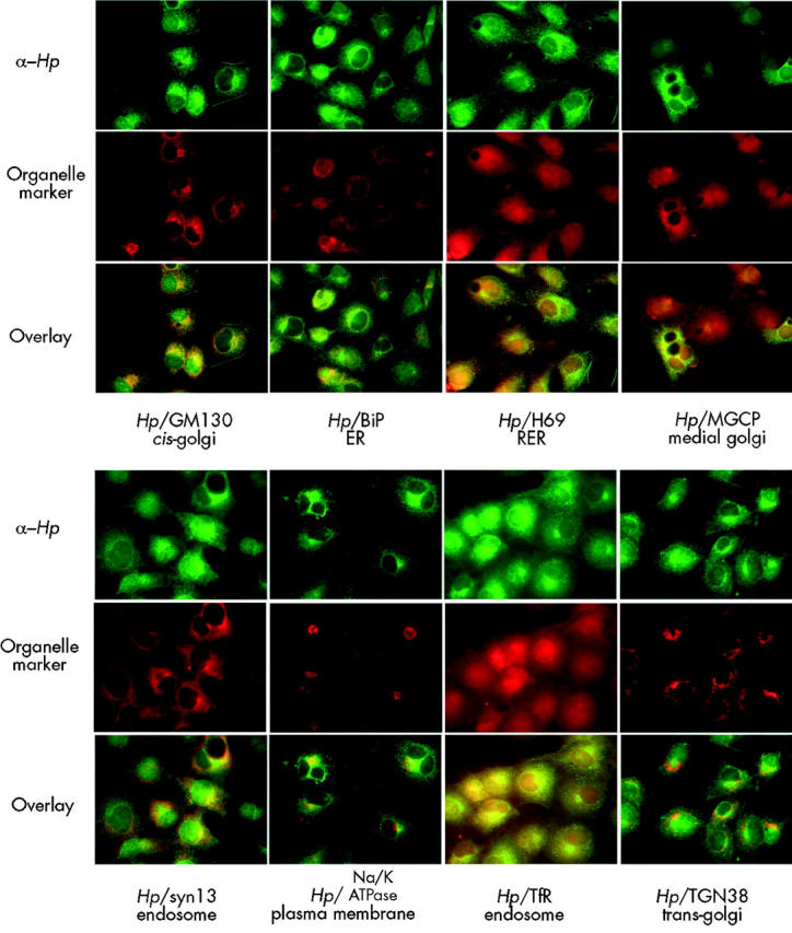
Colocalisation of hephaestin (Hp) with subcellular markers. Colocalisation of affinity purified antibodies to the C terminus of Hp (Hp1A) and antibodies to various subcellular markers in cultured CoS7 cells. Hp staining is represented in green and the markers (TfR, TGN38, GM130, BiP, H69, Na,K-ATPase, MGCP, and syntaxin 13) are shown in red. Yellow in the overlays indicates colocalisation. ER, endoplasmic reticulum; RER, rough endoplasmic reticulum; Tfr, transferrin receptor.