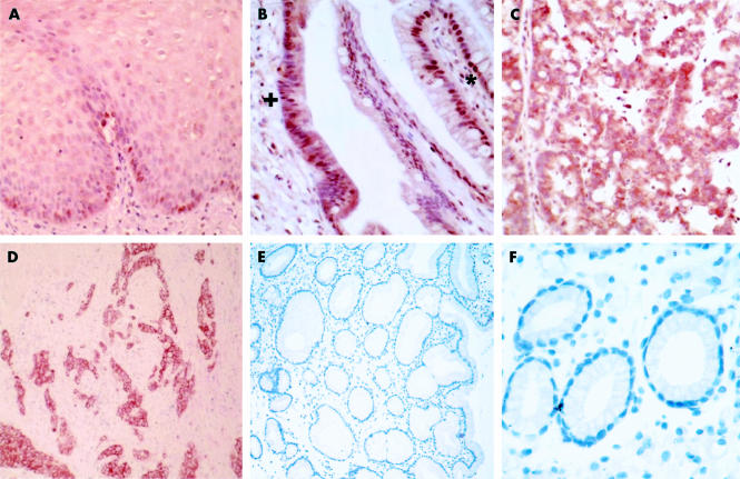
Figure 2.
Immunolocalisation of c-myc in the malignant progression of Barrett’s oesophagus (×40 objective), demonstrating localised nuclear staining in the basal layer of the squamous epithelium (A) and in benign metaplasia (B) (denoted by *), becoming cytoplasmic in dysplasia (B) (denoted by +). (C) High grade dysplasia exhibiting both nuclear and cytoplasmic c-myc immunoreactivity, which was further accentuated in adenocarcinoma (D). Adenocarcinoma at lower magnification (E) (×25 objective). No c-myc immunoreactivity was observed in gastric mucosa (F).

