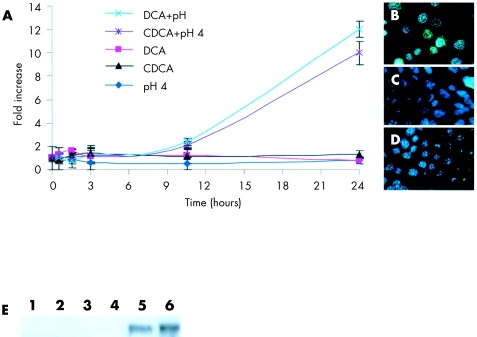
Figure 3.
C-myc mRNA levels in TE-7 cells over a 24 hour stimulation period (A). Induction of c-myc in cells stimulated with deoxycholic acid (DCA) at pH 4 or chenodeoxycholic acid (CDCA) at pH 4, but not in cells stimulated by DCA or CDCA at pH 7 or acidified (pH 4) growth media alone. Data are the mean (±2 SEM) of three experiments. Immunofluorescence of c-myc (FITC) in TE-7 cells stimulated by DCA at pH 4 or CDCA at pH 4 (B), DCA or CDCA at pH 7, or acidified growth media (pH 4) alone (C) and unstimulated cells (D) (×60 objective). (E) C-myc protein expression by western blotting. Lane 1, TE-7 cells cultured in growth media alone; lane 2, pH 4; lane 3, DCA (pH 7); lane 4, CDCA (pH 7); lane 5, DCA (pH 4); and lane 6, CDCA (pH 4). HeLa and Cos cell lysates were used as positive (+) and negative (−) controls, respectively. Cell lysates were normalised for equal protein loading by Coomassie blue staining.

