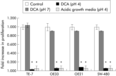
Figure 5.
Proliferation levels assessed in TE-7, SW-480, OE33, and OE21 cell lines following 24 hour exposure to deoxycholic acid (DCA) (pH 7), DCA (pH 4), acidic growth media (pH 4) compared with cells cultured in growth media alone (control). Data are the mean (±2 SEM) of three experiments. *Significant difference (p<0.05) between stimulated and unstimulated cells.

