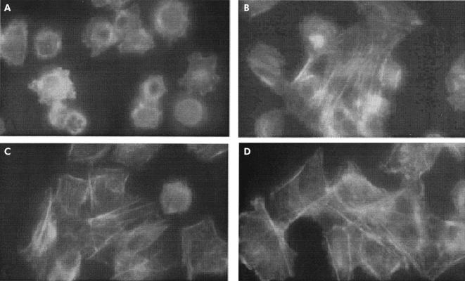
Figure 1.
Effect of gliadin (0.1 mg/ml) on IEC-6 cell cytoskeleton. (A) Fluorescence microscopy of gliadin exposed IEC-6 cells. Incubation for 15 minutes of cultured cells with gliadin caused a reorganisation of actin filaments characterised by redistribution to the cell subcortical compartment and subsequent cell rounding. A normal F-actin fluorescence pattern was observed when cells were exposed to similar concentrations of either zein, a protein from maize (B), or bovine serum albumin negative control (C). The gliadin effect on the actin cytoskeleton was reversible as two hours after withdrawal of gliadin from the culture medium the actin cytoskeleton returned to its basal state (D). Magnification: 40×.

