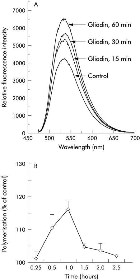
Figure 2.
F-actin quantitation by spectrofluorimetry in IEC-6 cells. (A) Gliadin (0.1 mg/ml) induced a time dependent increase in the cellular content of actin filaments, beginning as early as 15 minutes after exposure to the protein. Fluorescence was measured as relative fluorescence intensity units. (B) IEC-6 cells were exposed to gliadin 0.1 mg/ml at increasing time intervals, NBD-phallacidin extracted at the indicated time interval, and measured by spectrofluorimetry. The time profile of actin polymerisation showed a peak at 60 minutes. Actin polymerisation was expressed as per cent of control. n=4 for each time point.

