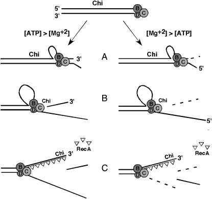
Figure 1.—
Two reactions of purified RecBCD enzyme at Chi, depending on the ATP:Mg2+ ratio. (Top) RecBCD binds to a dsDNA end, the RecB helicase being engaged with the 3′-end and the RecD helicase with the 5′-end. RecD is faster than RecB, and a ssDNA loop accumulates on the 3′ → 5′ (“top”) strand ahead of the RecB subunit (A). (Left) If the concentration of ATP is greater than that of Mg2+, RecBCD nuclease activity is low, and the enzyme nicks the top strand at Chi (B). (Right) If the concentration of Mg2+ is greater than that of ATP, RecBCD nuclease activity is high, and the enzyme degrades the top strand up to Chi (B), nicks the bottom strand, and degrades the bottom strand to the left of Chi (C). RecBCD loads RecA protein onto the top strand to the left of Chi (the “Chi tail”) (C). See Introduction and discussion for further description and references.

