
Figure 2.
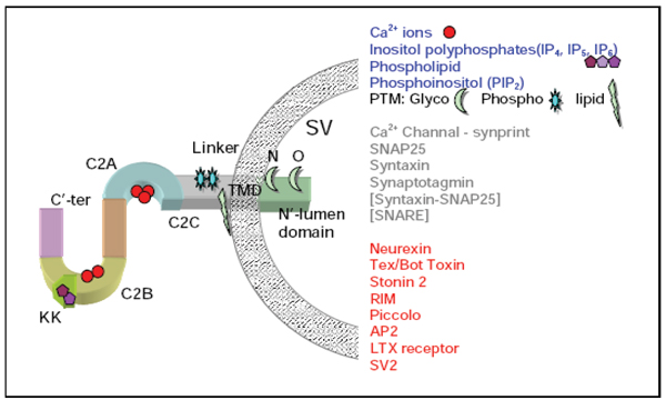
A schematic view of synaptotagmin1 (Syt1). Each functional domain is differently colored. Small molecules that interact with Syt1 (blue letters) and the post translational modifications (black letters) are listed. Proteins of the exocytotic apparatus and their unique combinations are marked in gray. Additional proteins that were shown to interact with Syt1 are shown in red. C2C is the linker sequence between the two C2 domains. Modification sites for N- and O-glycosylations are marked as N- and O-, respectively. TMD, transmembrane domain; KK, a stretch of basic residues that defines the binding site for inositol polyphosphates [30].