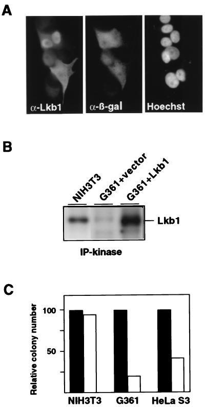
Figure 2.
Ectopic expression of Lkb1 in cells with impaired endogenous Lkb1 activity. (A) Subcellular localization of ectopic Lkb1 in G361 cells. Double immunofluorescence with anti-Lkb1 (Left) and anti-β-galactosidase (Center), indicating transfected cells. Nuclei were visualized by Hoechst staining (Right). (B) Lkb1 kinase activity in untransfected NIH 3T3 cells and G361 cells transfected with a Lkb1 encoding plasmid (G361 + Lkb1) or an empty vector (G361 + vector). Immunoprecipitation from cell lysates containing 400 μg of total protein and kinase activity assays were as performed as described in Fig. 1, except that samples were visualized by autoradiography. (C) G418-resistant colony formation by NIH 3T3, G361, and HeLa S3 cells after transfection with a vector control (solid bars) or with an Lkb1-expressing plasmid (open bars). For each cell line, the colony numbers obtained in the Lkb1 transfection were normalized to the vector transfection. A representative experiment is shown.

