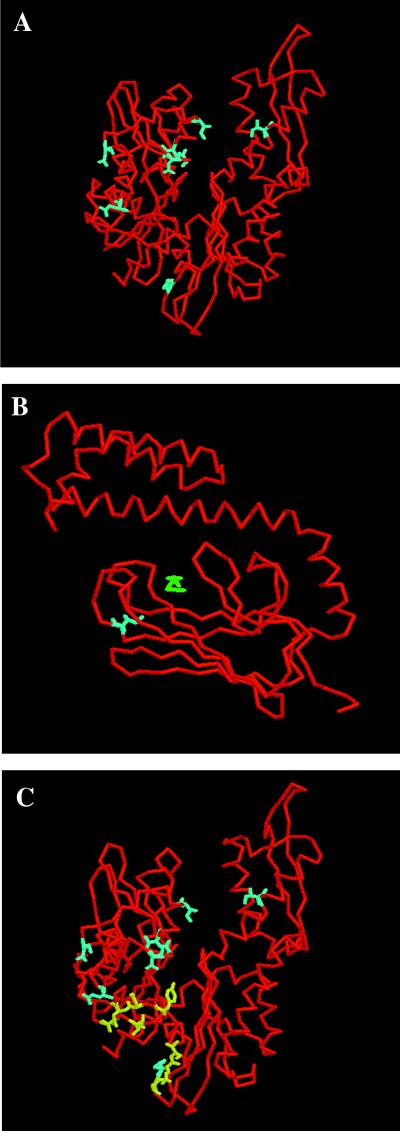
Figure 3.
Location of residues on DnaK crystal structures (PDB entries 1DKG and 1DKX). ATPase domain (A) is rotated 180° relative to the traditional view; peptide-binding domain (B). Residues described in this study are blue: Ssc1 D107 (DnaK D79), E109 (E81), V110 (V82), Q116 (I88), N126 (N98), Q180 (Q152), F239 (F216), L260 (I237), I485 (I462). Peptide substrate is green. Residues identified in other studies (C) are yellow: DnaK R167, I169, T215 (24); DnaK Y145, N147, D148, E217, V218 (25).

