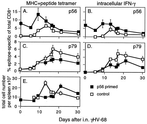
Figure 3.
The response in the spleen after respiratory γHV-68 challenge of mice primed with vacc-p56 and boosted with WSN-p56. The B6 mice were infected i.p. with vacc-p56 and i.n. with WSN-p56 1 month later. After another month, a few were sampled for analysis (day 0 time point), whereas a majority were challenged i.n. with γHV-68. The numbers of CD8+ T cells specific for the p56 (A and B) or p79 (C and D) epitopes were determined by staining with the p56Db (A) or p79Kb tetramers (C) or by in vitro stimulation with the p56 (B) or p79 (D) peptides in the presence of brefeldin A followed by staining for cytoplasmic IFN-γ. The kinetics of the splenomegaly that develops in these mice is illustrated in E.

