Figure 3.
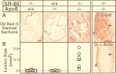
Effects of SR-BI gene disruption on atherosclerosis in apoE KO mice. Atherosclerosis in SR-BI−/− (n = 8, 4–6 weeks old), apoE−/− (n = 8, 5–7 weeks old), or SR-BI−/− apoE−/− (n = 7, 5–6 weeks old) female mice was analyzed in cryosections of aortic sinuses stained with oil red O and Mayer’s hematoxylin as described in Materials and Methods. (A) Representative sections through the aortic root region. (Bar = 200 μm.) (B) Sizes (cross-sectional areas) of oil red O-stained lesions in the aortic root region (see Materials and Methods). Average lesion areas (mm2 ± SD) for SR-BI−/−apoE−/−, apoE−/−, or SR-BI−/− mice, respectively, were as follows: 0.10 ± 0.07 (horizontal line), 0.002 ± 0.002, and 0.001 ± 0.002 (P = 0.0005). Also see Table 1. (C and D) High-magnification views of serial sections of plaque from the aortic sinus of a 7-week-old SR-BI/apoE double KO male, stained either with oil red O and Mayer’s hematoxylin (C) or with an anti-α actin antibody that recognizes smooth-muscle cells (D). The lumen is to the left of the plaque. The smooth-muscle wall (arrowheads) and cellular cap (arrows) are indicated. (Bar = 100 μm.)

