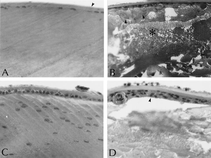
Figure 3.
Histology of lenses from clear or cataractous eyes. (A and C) Lenses from an estradiol-treated animal. (B and D) Lenses from a no-hormone animal. The eye of the estradiol-treated animal appears clear on gross examination, whereas the eye of the no-hormone-treated animal is opaque. (A) The anterior cortex of the estradiol-treated animal has a homogenous appearance and is covered by a lens capsule made of a thin epithelial cell layer and a normal, thick lens capsule (arrowhead). (B) The anterior cortex of the opaque eye is disrupted with the appearance of balloon cells (arrowhead) and areas of complete fiber degeneration and liquefaction (asterisk); the lens capsule is normal in appearance. (C and D) In the equatorial region, the clear lens (C) exhibits swelling of the nucleated fibers in the bow area and the fibers have a slight granular appearance. In the opaque lens (D), the fibers in the bow area are disrupted and the lens epithelium is hyperplastic (arrowhead). [Magnification = 250× (A, C, and D) and 125× (B).]

