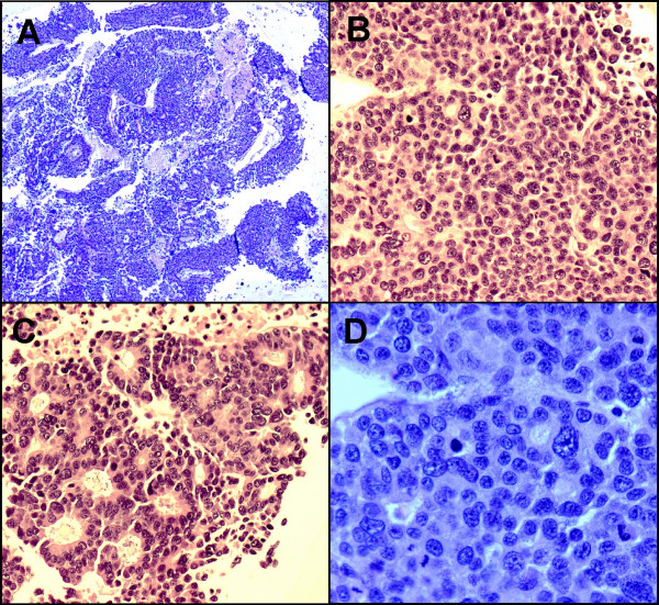
Figure 3.
A. On cell block, the tumor cells arranged in thick trabeculae, poorly formed glands or acini. Necrosis is evident. (H&E stain ×4); B. Solid pattern. (H&E stain ×20); C. Acinar pattern with uniform, eccentric located nuclei with abundant eosinophilic cytoplasm. (H&E stain ×20); D. Tumor cells with pleomorphic, centrally located nuclei, small to prominent nucleoli and scant to moderate amount of cytoplasm. Brisk and abnormal mitosis are evident. (H&E stain ×40)

