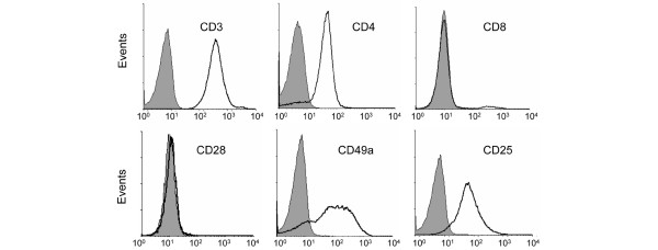
Figure 2.

Phenotypic analysis of type I collagen (CI)-responsive T-cell lines. T cells were expanded in vitro with autologous peripheral blood mononuclear cells, interleukin-2, and antigen (CI). The resultant T-cell lines that proliferated to CI were CD3+CD4+CD8-CD28-CD25+CD49a+. Grey shaded areas represent staining with the appropriate isotype control.
