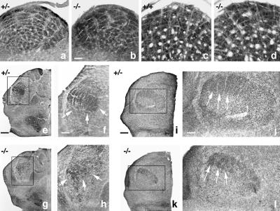
Figure 3.
(a +/− and b −/−) CO histochemistry of P7 trigeminal subnucleus interpolaris shows the brainstem whisker representation. (c +/+ and d −/−) CO histochemistry at P60 shows that the brainstem whisker representation persists in −/− mice. +/+, n = 2; +/−, n = 2, not shown; −/−, n = 2. (Bar = 100 μm.) (e and f +/−; g and h −/−) Horizontal section through left VB complex at P7 labeled by CO histochemistry of whisker representation (barreloids) in +/− mice (+/+ not shown). Higher magnification (f and h) of −/− mice shows that the boundaries of the VB thalamus are difficult to discern because of a much smaller area of intense CO labeling (arrows), suggesting that thalamic circuitry is relatively inactive. (e and g, Bar = 250 μm; f and h, Bar = 100 μm.) +/+, n = 2, not shown; +/−, n = 3; −/−, n = 2. (i–l) Horizontal oblique sections through the VB thalamus of P7 mice immunostained for 5HT-T show segregated barreloids in +/− and −/− thalamus. Higher magnification (j and l) shows that barreloid segregation in the −/− mouse (arrows) resembles +/+ (not shown) and +/−. (Bars = 100 μm.) +/+, n = 2; +/−, n = 3; −/−, n = 2.

