
Figure 1.
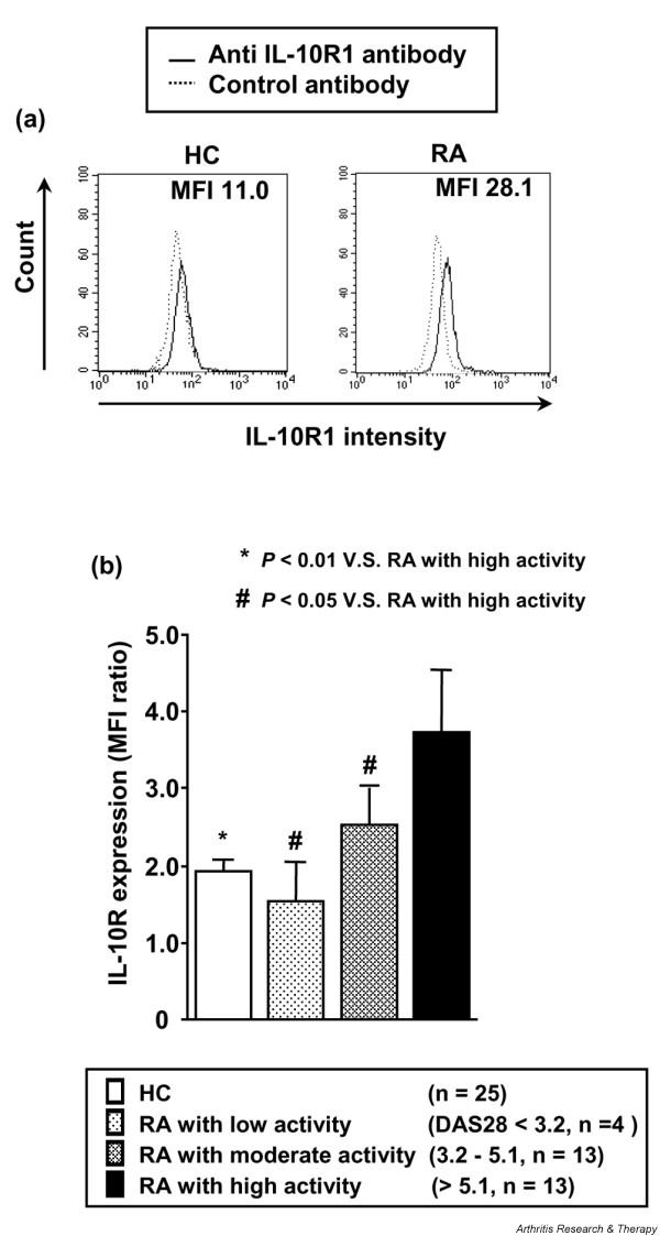
Expression of type 1 interleukin-10 receptor (IL-10R1) on monocytes from patients with rheumatoid arthritis (RA). (a) Representative histographic patterns of IL-10R1 expression on monocytes of patients with RA and healthy controls (HCs). Peripheral blood mononuclear cells were stained with mouse immunoglobulin (Ig) G1 anti-IL10R1 antibody or isotype-matched control antibody, followed by fluorescein isothiocyanate-conjugated goat anti-mouse IgG1 antibody. Flow cytometric analysis of IL-10R1 expression was performed by gating on monocytes according to light scatter profile. (b) Increased IL-10R1 expression on monocytes from RA patients with active disease. Patients with RA were divided into groups with low, moderate, and high disease activity according to the disease activity score 28 (DAS28) system. The intensity of cell surface IL-10R1 expression on monocytes from RA patients and HCs was expressed by the ratio of the mean fluorescence intensity (MFI) of staining with anti-IL-10R1 antibody to the MFI of control antibody. Values are the mean ± standard error of the mean. n, number of samples tested.