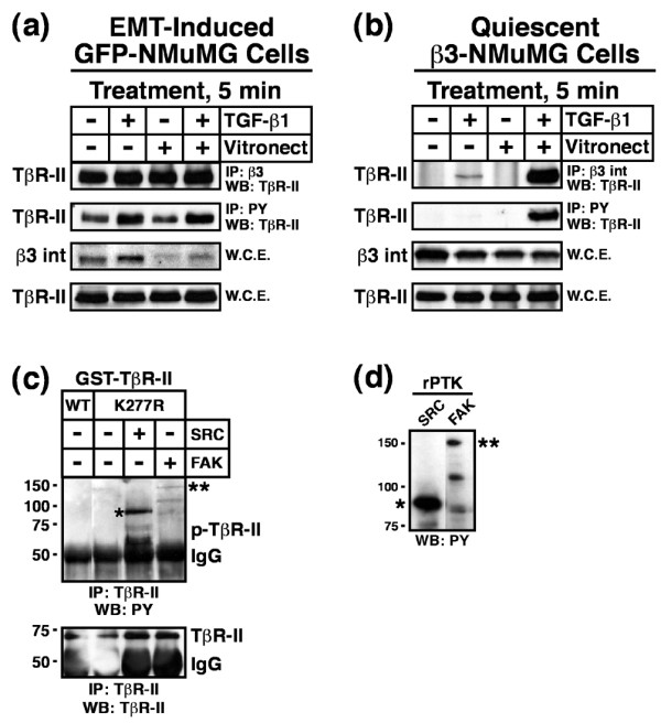
Figure 6.

Dual receptor activation induces Src mediated TβR-II tyrosine phosphorylation. (a) GFP-expressing NMuMG cells were stimulated with TGF-β1 (5 ng/ml) for 36 hours to induce β3 integrin expression and EMT. Afterward, the cells were dissociated and re-plated either onto plastic or vitronectin-coated wells in the absence or presence of TGF-β1 (5 ng/ml) for 5 min. Afterward, detergent-solubilized whole cell extracts were prepared (1 mg/tube) and subsequently immunoprecipitated with anti-phosphotyrosine or anti-β3 integrin antibodies as indicated. The presence of TβR-II in precipitated immunocomplexes was determined by Western blotting with anti-TβR-II antibodies. Differences in protein loading were monitored by immunoblotting whole cell extracts (50 mg/lane) for β3 integrin and TβR-II. Shown are representative immunoblots from a single experiment that was repeated twice with similar results. (b) Resting WT β3 integrin-expressing NMuMG cells were dissociated and re-plated as above. Afterward, TβR-II tyrosine phosphorylation and complex formation with β3 integrin was determined by immunoblotting as above. Shown are representative immunoblots from a single experiment that was repeated three times with identical results. (c) Active recombinant Src (1 unit/reaction) or FAK (0.2 μg/reaction) kinases were added to protein kinase reaction mixtures containing 1 μg/tube of either kinase active (WT) or inactive (K277R) GST-TβR-II. Phosphorylation reactions were stopped after 30 min and the phosphorylation status of TβR-II was determined by immunoprecipitation of diluted protein kinase reaction mixtures with anti-TβR-II antibodies. Afterward, immobilized immunocomplexes were probed sequentially with anti-phosphotyrosine antibodies, followed by anti-TβR-II antibodies as indicated (*autophosphorylated recombinant Src; **autophosphorylated recombinant FAK). Data are from a representative experiment that was repeated three times with similar results. (d) Recombinant Src and FAK were incubated in protein kinase assay buffer for 30 min at 30°C. The data show the autophosphorylation on tyrosine residues of recombinant Src (*) and FAK (**) as determined by immunoblotting with anti-phosphotyrosine antibodies. Data are from a representative experiment that was repeated at least three times with similar results. EMT, epithelial-mesenchymal transitions; FAK, focal adhesion kinase; GST, glutathione S-transferase; TβR, TGF-β receptor; TGF, transforming growth factor; WCE, whole cell extracts; WT, wild type.
