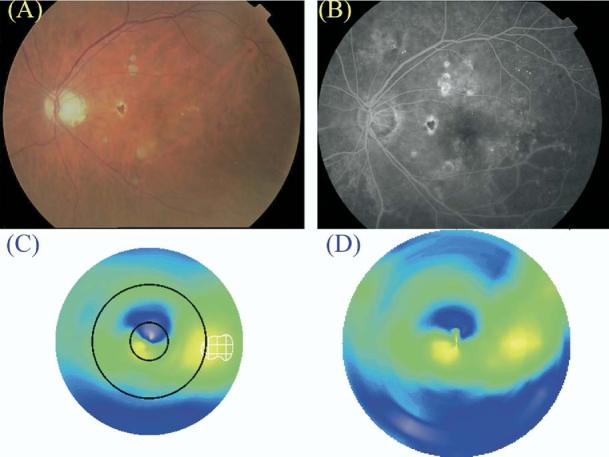
Figure 8.

Patient diagnosed to have clinically significant macular edema (CSME) 1 by clinical examination (A, color fundus photograph; B, late fluorescein angiogram frame) but not by Macular Grid 5 (C, MG5 thickness map) or Fast Macular Thickness Map (D, FMTM) protocols. The map of edema as identified by the MG5 algorithms is delineated by the white checkered zone in C. Note that retinal thickening (compared with the normal reference) was present in the central circle of the MG5 map but did not meet the threshold level defined in this study.
