


Figure 2.
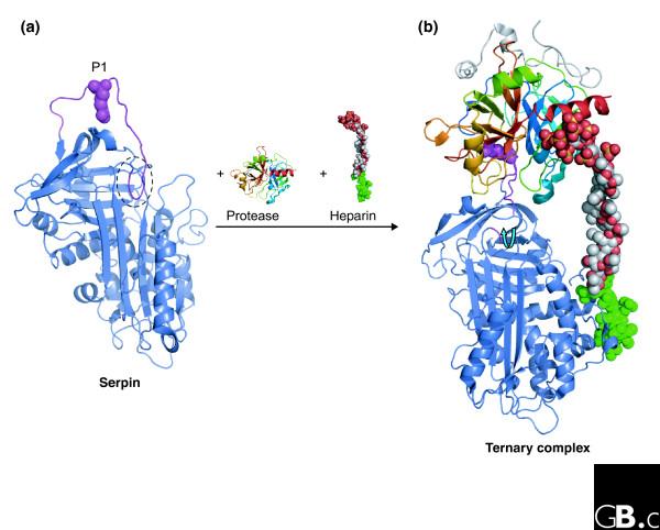
Modulation of serpin conformation by cofactors. (a) The structure of native SERPINC1 (PDB code 2ANT) [95]. The partial insertion of the RCL (two residues) into the top of β-sheet A is circled, and the position of the P1 residue is shown (magenta spheres). (b) The structure of the ternary complex between SERPINC1, inactive thrombin (the Ser195Ala mutant) and a synthetic long-chain heparin construct (PDB code 1TB6) [43]. A specific high-affinity pentasaccharide (green) on the heparin interacts with the heparin-binding site on SERPINC1 (on and around helix hD) and promotes expulsion of the RCL (blue arrow) and rearrangement of the P1 residue (magenta spheres).