Figure 5.
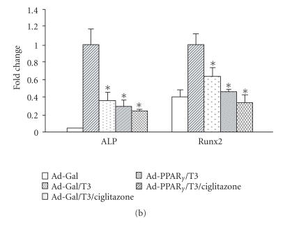
Inhibition of T3-induced hypertrophy and mineralization in the growth plate cells by PPARγ overexpression or ciglitazone treatment. (a) Growth plate cells were treated with T3 (100 ng/mL) in the presence or absence of ciglitazone (5 μM) for 10 days. Alkaline phosphatase (ALP) staining was used as a marker of terminal differentiation of growth plate chondrocytes. Positive stainings were colored in dark blue. Negative-stained background was colored in light green. (b) Quantitative PCR analysis of ALP and Runx2 genes 4 days after growth plate cells were infected with Ad-PPARγ or treated with ciglitazone. Ad-Gal-infected cells without T3 or ciglitazone treatment were used as controls. Gene expression levels were normalized with respect to endogenous 18S rRNA. * P < .05 versus the expression in the Ad-Gal-infected cells with T3-treatment.


