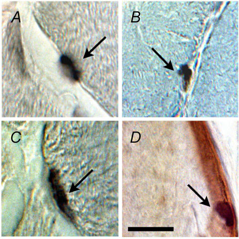
Figure 1.
Photomicrographs of representative immunohistochemical stains of M-cadherin (A), BrdU (B), myogenin (C), and laminin and haematoxylin (D). These stains were used to identify satellite cells (A; arrow), proliferating satellite cells (B; arrow), terminally differentiating satellite cell progeny (C; arrow) and intrafibre muscle nuclei (D; arrow). Scale bar represents 40 μm.

