
Figure 6.
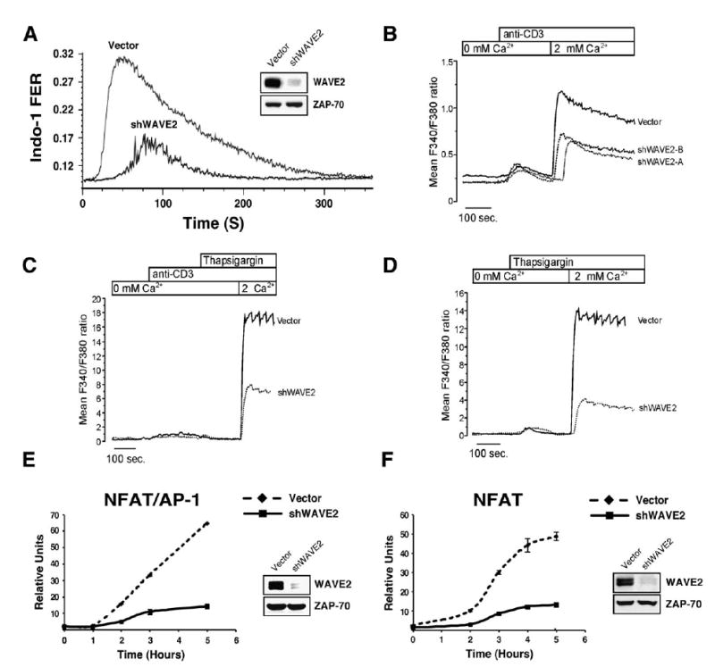
WAVE2 Regulates CRAC Channel Activation Leading to NFAT-Mediated Gene Transcription
(A) Jurkat T cells were transfected with vector control or shWAVE2 suppression vector. After 72 hr, calcium mobilization in response to OKT3/ goat anti-mouse was measured using Indo-1 staining and flow cytometry.
(B) Jurkat T cells were transfected as in (A). After 72 hr, calcium measurements were performed using single cell fluorescence ratio (Fura-2 imaging) of GFP positive control and WAVE2 suppressed Jurkat cells. In calcium-free bath solution, increases in Fura-2 ratio reflect Ca2+ release from intracellular stores. Extracellular Ca2+ entry via activated CRAC channels was subsequently assessed by reintroduction of extracellular calcium (2 mM Ca2+). Shown are representative examples of three independent experiments for each condition and each trace represents the average response of at least 100 cells in the recording chamber.
(C and D) Cells were prepared and analyzed as in (B) and stimulated as indicated above the graphs.
(E and F) Jurkat T cells were transfected with vector control or shWAVE2 suppression vector and either a (E) NFAT/AP-1 or (F) NFAT luciferase reporter. 72 hr post transfection, cells were stimulated with OKT3 and anti-CD28 for 2, 3, 4, and 5 hr. Luciferase activity was measured at each time point and normalized to TK-Renilla readings. Shown are representative examples of three independent experiments for each.