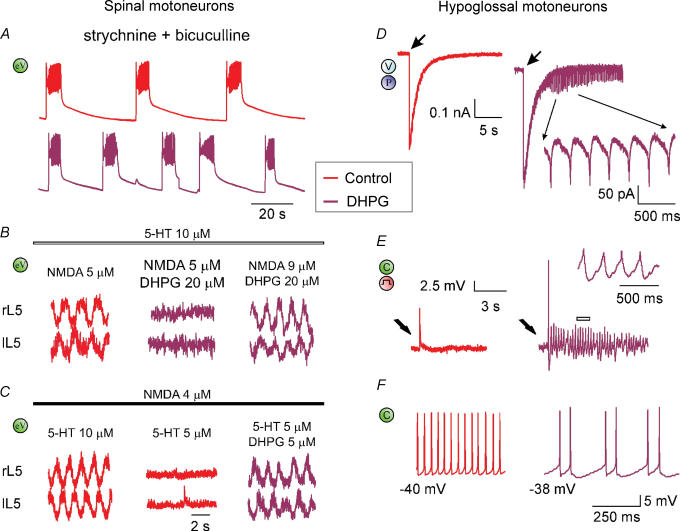
Figure 3. Network actions of group I mGluR activation.
A, disinhibited bursting evoked by block of synaptic inhibition (red trace; 1 μ m strychnine plus 20 μ m bicuculline application) is accelerated by DHPG (5 μ m; crimson trace) which significantly reduces burst duration and periodicity. Because disinhibited bursting is believed to originate from the rhythmic discharges by CPG, this effect suggests activation of CPG interneurons by DHPG. eV (green), extracellular DC recording from VR. B, fictive locomotor patterns induced by co-application of NMDA and 5-HT is depressed by 20 μ m DHPG and restored by increasing the NMDA concentration to 9 μ m. C, conversely, fictive locomotor patterns brought below threshold by decreasing the 5-HT concentration are restored by adding a small (5 μ m) dose of DHPG. Data are reproduced, with permission, from Taccola et al. (2004b). D, fast inward current recorded (under voltage clamp, V) from single HM induced by puffer application (P) of AMPA is followed by rhythmic oscillations when occurring in coincidence with a concentration of DHPG (5 μ m) subthreshold for oscillations. E, electrically evoked EPSP (by stimulating the DMRC, see step symbol in red) recorded under current clamp (C, green) in control solution (red trace) generates spike and oscillations (crimson trace; see also inset at faster time scale for record corresponding to the open bar) when repeated in the presence of DHPG (5 μ m). F, comparison of two HMs (left and right) at similar membrane potential in the presence of DHPG (20 μ m). The one on the left (red) produces high-frequency firing without oscillations, while the one on the right (crimson) generates oscillations with lower firing rate. Data reproduced with permission from Sharifullina et al. (2005).

