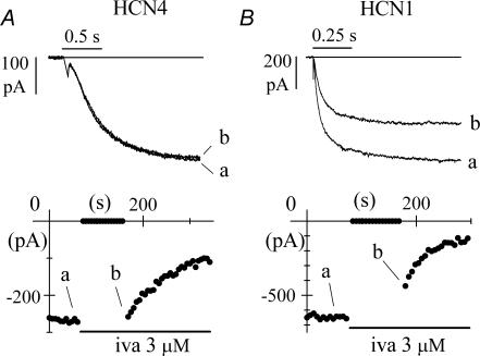
Figure 2. Block by ivabradine requires open HCN4, but does not require open HCN1 channels.
A, time course of IHCN4 amplitude at −100 mV during a standard pulsing protocol (−100 mV, 1.8 s; +5 mV, 0.45 s). The cell was rested at −35 mV for the first 90 s of ivabradine (3 μ m) perfusion before resuming the pulsing protocol. During the period at −35 mV no current reduction was observed. B, time course of IHCN1 amplitude at −100 mV during an identical protocol. In this case, during the time spent at −35 mV a significant current reduction was indeed observed, indicating partial block development. In A and B, sample traces recorded at various times as indicated.

