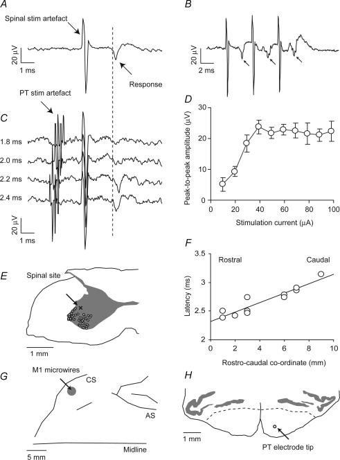
Figure 1. Identification of antidromic cortical potential evoked by intraspinal stimulation.
A, field potential recorded differentially between two microwire electrodes in primary motor cortex following a spinal stimulus of 60 μA (average of 120 sweeps). B, cortical field (indicated by arrows) followed each of three stimuli at 250 Hz (average of 60 sweeps). C, a collision test established that the field is antidromic. When pyramidal tract (PT) stimulation at 500 μA preceded the spinal stimulus by 2.0 ms or less, the cortical response was abolished. With an interval of 2.2 ms or more, the field appeared (40 sweeps per interval). For clarity, the averaged response to PT stimulation alone, which exhibits a large antidromic field, has been subtracted from each trace. A small artefact remains due to incomplete cancellation of the rising and falling phases of the stimulation artefact. D, peak-to-peak amplitude of antidromic cortical response to different intensities of spinal stimulation. E, post-mortem localization of lesion made at the C7 level from which an antidromic cortical response was elicited. The lesion site (marked x) was within the grey matter on the mediodorsal edge of the motoneuron territory (large cell bodies, marked ^). F, latency of antidromic response onset as a function of rostrocaudal location of stimulus sites in the recording chamber. G, location of cortical microwire implant relative to the central sulcus (CS) and arcuate sulcus (AS) based on post-mortem photographs. H, transverse section through the brainstem at A2, showing location of anterior PT electrode and grey matter of the olive. Dashed lines indicate the approximate border of the PT. The tip of the second electrode was 5 mm posterior.

