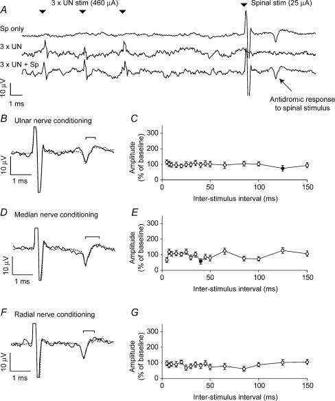
Figure 3. Absence of conditioning effects from peripheral nerve stimulation.
A, the three traces show averaged responses to spinal stimulation (25 μA), a train of stimuli (3 × 460 μA at 300 Hz) delivered to the ulnar nerve, and spinal stimulation preceded by ulnar nerve stimulation with an interstimulus interval of 10 ms. B, expanded plot comparing the response to spinal stimulation alone (dashed line) with the response following ulnar nerve conditioning (continuous line). No facilitation of the response was seen. C, modulation of spinally conditioned response for different interstimulus intervals. D and E, comparable plots for median nerve stimulation (3 × 520 μA at 300 Hz). F and G, comparable plots for radial nerve stimulation (3 × 5.2 mA at 300 Hz).

