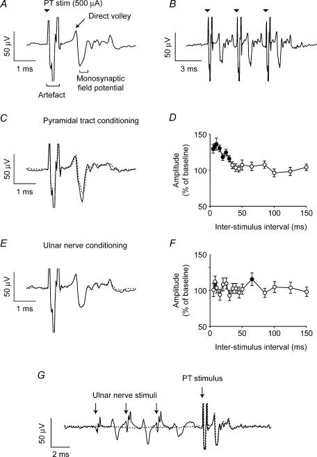
Figure 7. Conditioning of monosynaptic spinal field potential evoked by PT stimulation.
A, spinal cord recording of the field evoked by PT stimulation at 500 μA (average of 120 sweeps). B, monosynaptic field was enhanced following each of a train of three PT stimuli at 300 Hz (average of 70 sweeps). C, monosynaptic field was facilitated by paired PT stimulation with an interstimulus interval of 15 ms (continuous line) relative to a single stimulus (dashed line). D, percentage modulation of monosynaptic field following paired-pulse PT stimulation with different interstimulus intervals. E, monosynaptic field was unaffected by preceding ulnar nerve stimulation (3 × 400 μA at 300 Hz). F, modulation of monosynaptic field following a conditioning stimulus to the ulnar nerve with different interstimulus intervals. G, spinal field potentials evoked by stimulation of PT, without (dashed) and with (continuous) the ulnar nerve (3 × 400 μA at 300 Hz). Stimulus artefacts are indicated by arrows. Average of 40 sweeps.

