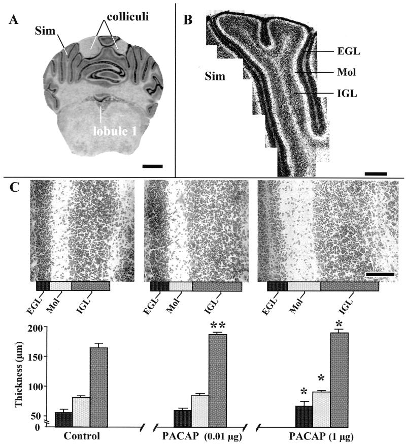
Figure 2.
Effects of graded doses of PACAP on the histogenesis of the Sim in P12 rat cerebella. (A) Typical Cresyl Violet staining of a rat cerebellum slice at the caudal extremity of the colliculi and the rostral extremity of lobule 1, showing the type of section used for measurements with the confocal laser-scanning microscope. (Bar = 1.4 mm.) (B) Higher magnification of a Sim showing the series of fields that were used for the measurement of the thickness of the EGL, the molecular layer (Mol), and the IGL, as well as for the measurement of the density of granule cells in the molecular layer and IGL. (Bar = 400 μm.) (C) Quantification of the thickness of the EGL, molecular layer, and IGL from the Sim of P12 rats. (Bar = 100 μm.) Each value represents the mean ± SEM of a representative experiment performed in triplicate. ∗, P < 0.05 vs. control; ∗∗, P < 0.01 vs. control.

