
Figure 8. Leptin had no effect on spontaneous IPSC frequency (sIPSC).
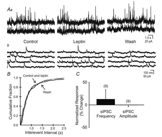
Aa, spontaneous IPSCs observed in this unidentified neurone before, during and after superfusion of leptin (100 nm; Vm = −15 mV). b, expanded portions of corresponding traces in A. B, cumulative fraction plot shows no significant change in sIPSC frequency in response to leptin (P > 0.05). C, percentage change of sIPSC frequency and amplitude in response to leptin application (P > 0.05).