
Figure 8. Distribution of the impact of charge mutants on the three-dimensional structure of hERG S5P α-helix.
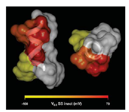
Orthogonal views of a space filling model of the hERG S5P α-helix, based on NMR structure of S5P linker (PDB: accession number 1JUL), with side chains colour-coded according to the effect lysine mutants have on the V0.5 of steady-state inactivation. Hydrophobic residues are shown in white.