
Figure 6. Effects of modulators of PKA, PKC and G-protein activity on IKs.
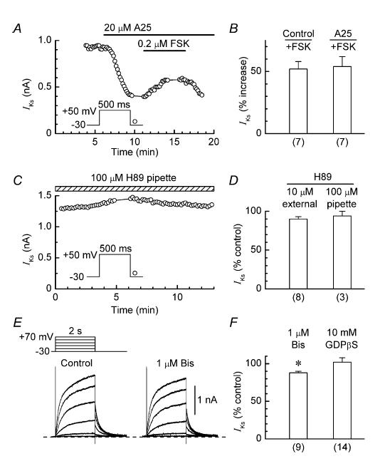
A, stimulation of IKs by 0.2 μm forskolin (FSK) in a myocyte pretreated with 20 μm tyrphostin A25 (A25). B, comparison of the stimulatory effects of 0.2 μm forskolin on IKs in control and tyrphostin-pretreated myocytes. C, lack of effect of H89 (100 μm) in the pipette solution on IKs in a representative myocyte. D, summary of the results obtained with myocytes that were treated with external H89 (10 μm) or internal H89 (100 μm) for ∼12 min. For evaluation of the effects of internal H89, the average amplitude of the IKs tail during the first minute after patch formation was taken as the control amplitude. E, records obtained from a myocyte before and 20 min after addition of 1 μm bisindolylmaleimide I (Bis). F, summary of the effects of treatments for 10–20 min with external 1 μm bisindolylmaleimide I and internal 10 mm GDPβS on the amplitude of the IKs tail; *P < 0.05. Number of myocytes is shown in parentheses.