
Figure 5. Lack of hypoxia sensitivity of Kv1.3 current in JCaM1.6 cells.
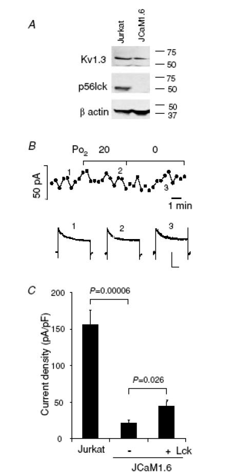
A, lack of Lck expression in JCaM1.6 cells. Immunoblot analysis of Kv1.3α subunit and Lck proteins in Jurkat (lane 1) and JCaM1.6 cells (lane 2). β-Actin was used as a constitutive protein in these samples. B, time course of Kv1.3 current amplitude measured in normoxia and during exposure to progressively more severe hypoxia (top). The continuous lines indicate the time of exposure to hypoxia. The Kv1.3 current recordings corresponding to the time-points marked by numbers are shown in the bottom panel. Scale bars correspond to 200 ms and 100 pA. Kv1.3 currents were elicited by step depolarizing steps from −80 mV HP to +50 mV, every 30 s. C, introduction of Lck into JCaM1.6 cells increases the Kv1.3 current amplitude. Kv1.3 current density, measured under whole cell patch-clamp configuration, is significantly lower in JCAM1.6 cells than in Jurkat cells (n = 10 in Jurkat and n = 9 in JCaM1.6). Kv1.3 currents were elicited as described in panel B. Introducing 1 U ml−1 active Lck into the cells by dialysing through the patch pipette significantly increases the current density in JCaM1.6 cells (n = 11).