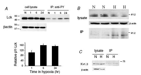
Figure 8. Activation of Lck by prolonged hypoxia and tyrosine phosphorylation of Kv1.3 channels.

A, T lymphocytes were exposed to normoxia and hypoxia (8 mmHg) for 1, 6 and 24 h. Cell lysates containing equal amount of total proteins were immunoprecipitated with rabbit polyclonal anti-phosphotyrosine antibody and the immunoprecipitated samples were analysed by Western blotting using rabbit polyclonal anti-Lck antibody. β-Actin was used as loading control. Hypoxia induced an increase in tyrosine phosphorylated Lck level after 6 and 24 h exposure. The lower panel shows the average densitometric values of tyrosine phosphorylated Lck after 1, 6 and 24 h exposure to hypoxia. Densitometric values of the immunoprecipitated bands were normalized to the corresponding values in the crude protein blots and were expressed as a percentage of the normoxic value. n = 6 donors for 1 h, and n = 2 donors for 6 and 24 h. B, Jurkat cells were exposed to normoxia and hypoxia (PO2 of 8 mmHg) for 24 h. Cell lysates containing equal amount of total proteins were immunoprecipitated with rabbit polyclonal anti-phosphotyrosine antibody. The cell lysate and the immunoprecipitated samples (IP) were analysed by Western blotting using rabbit polyclonal anti-Kv1.3 antibody. β-Actin was used as loading control. Hypoxia induced an increase in tyrosine phosphorylated Kv1.3 level. C, experiments similar to those described in A were conducted after maintaining Jurkat cells in either normoxia or hypoxia (8 mmHg) for 15 min. This blot is representative of 2 separate experiments.
