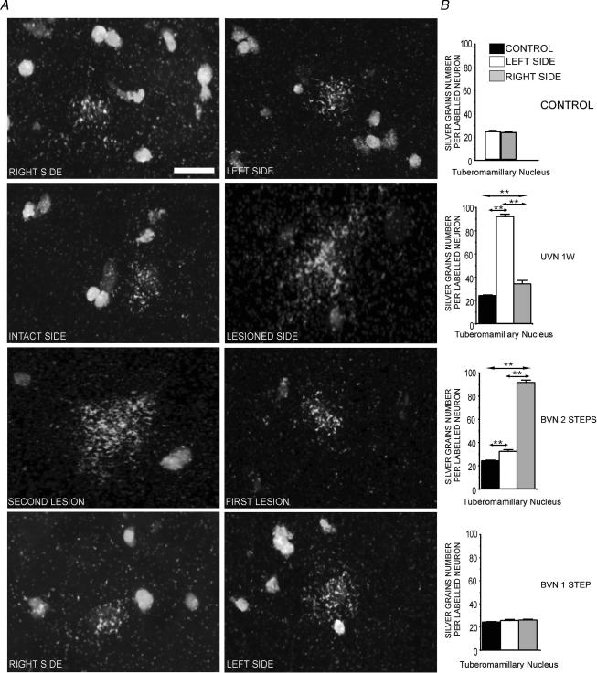
Figure 2. Changes in the number of silver grains per HDC radiolabelled neuron in the TM nuclei after vestibular lesion.
A, high magnification photomicrographs from emulsion autoradiograms showing hybridization of cRNA specific for histidine decarboxylase mRNAs to neurons in the tuberomammillary nucleus (TM), illustrating the number of silver grains per labelled neuron in the tuberomammillary nuclei (TMN) on the right and left sides of a representative control cat. One week after unilateral vestibular neurectomy (UVN), the number of silver grains on the intact side was lower than that on the lesioned side. In the two-step bilateral vestibular neurectomized cats (BVN), mirror images were obtained with an increased labelling on the side of the second lesion as compared with the TMN on the side of the first lesion. The number of silver grains per labelled neuron remained unchanged in the one-step BVN cats. Scale bar, 10 μm. B, quantitative analysis of the effects of unilateral vestibular neurectomy (UVN) and bilateral vestibular neurectomy (BVN) performed in one or two steps on the number of silver grains per radiolabelled neurons in the tuberomammillary nuclei. Data are expressed as mean values (±s.e.m.). The values recorded on the right (grey bars) and left (white bars) sides are given separately for all the vestibular lesioned cats. The data from both sides were pooled for the controls (black bars) for a direct comparison with the subgroups of cats killed 1 week after a left unilateral vestibular neurectomy, and with the two-step and the one-step bilateral vestibular neurectomized cats. The bar graphs correspond to those of Fig. 1B for the UVN cats examined at 1 week, and the one- and two-step BVN cats. **P < 0.0001. UVN: unilateral vestibular neurectomy; BVN: bilateral vestibular neurectomy; 1 W: 1 week.

