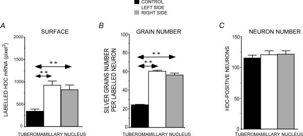
Figure 3. Effects of thioperamide treatment on HDC mRNA expression in the tuberomammillary nuclei.
Quantification of the HDC mRNA labelled surface (A), silver grain number per HDC labelled neuron (B), and the number of HDC radiolabelled neurons (C) on the right (grey bars) and left (white bars) TM nucleus of thioperamide-treated cats compared to the control cats (black bars). Note that the HDC mRNA labelled surface and the number of silver grains per HDC-labelled neurons are bilaterally and significantly increased in the TM nuclei of thioperamide-treated cats. By contrast, the number of radiolabelled neurons in the TM nuclei is not affected by the thioperamide treatment. **P < 0.0001. HDC: histidine decarboxylase.

