Figure 2.
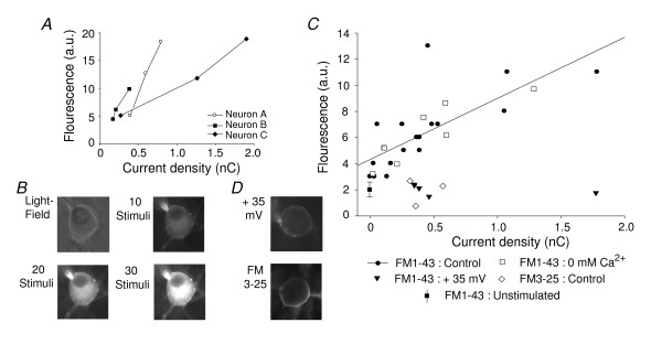
Cytoplasmic accumulation of FM1-43 via permeation through MS ion channels. Using the perforated patch configuration allowed dye accumulation in the cytoplasm to be measured. (A) Cytoplasmic fluorescence through FM1-43 uptake increased in 3 neurons after 10, 20 and 30 mechanical stimuli; shown is Intensity of fluorescent labelling against cumulative charge transfer. (B) Example images of a neuron (light field image, top left) following 10, 20 and 30 mechanical stimuli. N.B. the dye does not enter the nucleus. (C) Accumulation of FM1-43 is dependent on MS channel activity. In control conditions (standard external solution, membrane potential; -70 mV, ● solid line) fluorescent intensity is correlated with the amount of channel activity (as total charge transfer)(n = 18, Spearman's ranked order, r = 0.83, P < 0.001, fit: solid line). Removal of external Ca2+ (□) had no apparent effect on dye uptake (n = 8, Spearman's ranked order, r = 0.83, P < 0.001, fit dotted line, partly occluded). Neither application of FM1-43 when the neuron was held at +35 mV (n = 5, ▼) nor application of FM3-25 (at -70 mV, n = 3, ◇) resulted in significant cytoplasmic fluorescence. Also shown is average background labelling after FM1-43 exposure in the absence of mechanical stimulation (n = 10, standard deviation indicated, ■). D. Examples of neurons stimulated in FM1-43 at +35 mV (top) and in FM3-25 at -70 mV (bottom).

