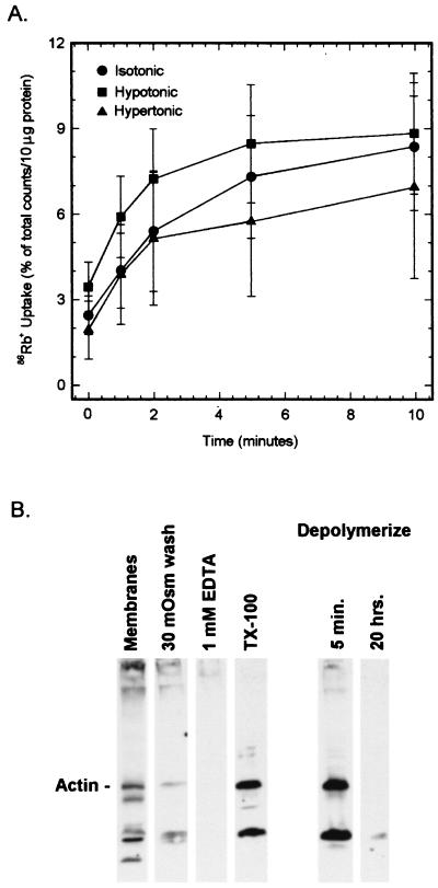
Figure 6.
(A) Blocking the volume regulatory response by depolymerization of actin. Vesicles were incubated overnight under conditions that promote the depolymerization of actin (see Methods). The vesicles then were equilibrated with a 410 mOsm solution and were assayed for 86Rb+ uptake as described in Fig. 1. Note that the higher specific channel activity, compared with that shown in previous figures, is likely attributable to loss of protein (e.g., cytoskeletal proteins) in the depolymerization procedure. (B) Immunoblot of actin in the basolateral membrane vesicles after depolymerization. Vesicles were treated as described in Methods under various conditions that are known to disrupt the cytoskeleton. The depolymerized vesicles were run in a parallel experiment to the transport assay shown in A.

