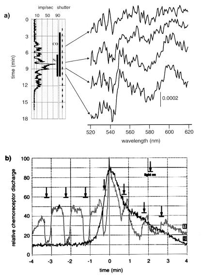
Figure 5.
(a) Chemoreceptor discharge (Left) and cytochrome redox state (Right) under the influence of CO in normoxia leads to discharge increase without light absorption changes (first spectrum trace). Hypoxia (N2) produces discharge increase with typically reduced cytochrome spectra. Short and repeated transilluminations of carotid body tissue, for recording absorption spectra (small bars in shutter), lead to discharge inhibition during normoxia and excitation during hypoxia. (b) Chemoreceptor discharge under normoxic and hypoxic CO applications. (Reference curve 1) Effect of CO as percentage of peak chemoreceptor activity induced by 4-min hypoxia (reference curve 2). Mean of 10 carotid bodies. CO application during normoxia (left side of time zero) lead to excitation, which is eliminated during transient transilluminations (vertical arrows). CO application during hypoxia (right side of time zero) leads to discharge inhibition, which is reversed during transillumination.

