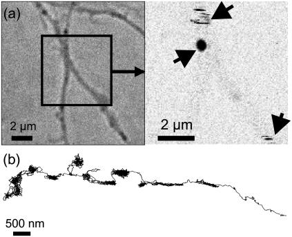
FIGURE 5.
White light image (a, left) and LISNA image (a, right) of a live neuron labeled with gold NPs. LISNA image exhibits signals from two moving (strips) and one stationary (spot) GluR2 receptors labeled with gold NPs (arrows). (b) Trajectory of an individual 5-nm gold NP (>5 min, 9158 data points) acquired at video rate on a live neuron (see also a movie in Supplementary Material).

