Fig. 1.
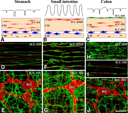
Overview of the types of ICC in the gastrointestinal tract. A–C: Schematic representations of ICC in the stomach (A), small intestine (B) and colon (C). ICC-MY (blue) are located between circular (CM) and longitudinal (LM) muscle layers. ICC-IM (red) and ICC-DMP (red) are located within circular and longitudinal muscles. ICC-SM (green) are located at the submucosal surface of the circular muscle layer in the colon. D–J: ICC and nerve networks in the murine stomach (D, E), small intestine (F, G) and colon (H–J). ICC and nerves in whole mount preparations are demonstrated by immunohistochemistry for Kit (ICC marker, green) and PGP9.5 (pan-neuronal marker, red), respectively. ICC-MY with a multipolar shape are associated with myenteric ganglia (Auerbach’s ganglia) (MG). ICC-IM in the stomach and colon have a bipolar shape and are associated with intramuscular nerve fibers. ICC-DMP in the small intestine are associated with deep muscular plexus. ICC-SM are multipolar cells having many thin processes. Bar=100 µm.

