
Fig. 2.
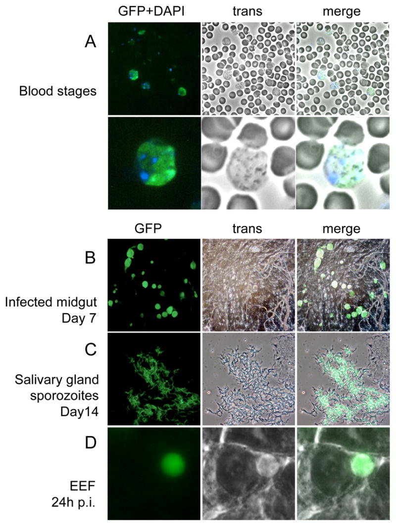
Fluorescence of PyGFP in different life stages. (A) blood stage parasites stained with DAPI, lower panel shows higher magnification; (B) infected midgut at day 7 post feeding; (C) sporozoites in salivary gland; (D) mature liver stage 24 h post hepatocyte invasion. Fluorescence, bright field and merged images of the same microscopic field are shown in each line.