
Figure 4.
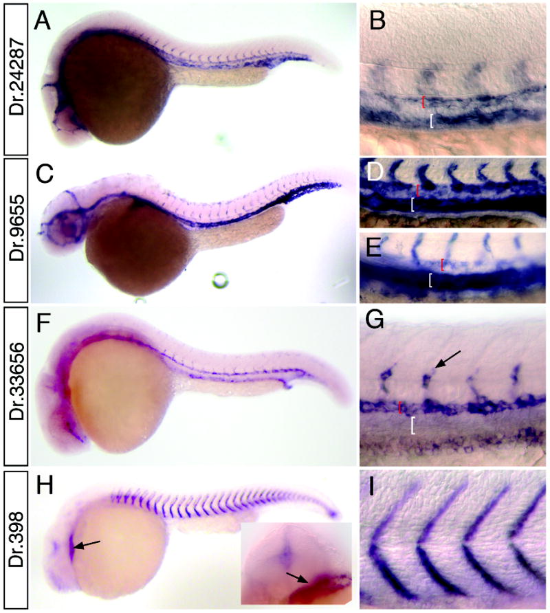
Whole mount in situ hybridization analysis of cardiovascular markers. A, B. Dr.24287 at 24 hpf; box indicates magnified view in B. B. Black bracket indicates dorsal aorta, white bracket shows posterior cardinal vein. C-E. Dr. 9655. C. Embryo at 30 hpf. D. Magnified view of dorsal aorta (red bracket) and posterior cardinal vein (white bracket) at 24 hpf; E. Dorsal aorta (red bracket) and posterior cardinal vein (white bracket) at 30 hpf. F, G. Dr.33656. G. expression in segmental arteries (black arrow) and dorsal aorta (red bracket); expression is absent from posterior cardinal vein (white bracket). H, I. Dr.398 at 24 hpf. H. Arrow indicated expression in heart; inset head-on view showing heart expression (arrow). I. Magnified view of embryo in H.