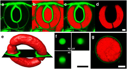
FIGURE 1.
3D reconstruction. Preprocessing of image stacks and evaluation of the reconstruction on simple objects. To the original data (a), a threshold was set, and pixels, with a value equivalent to the threshold (b, red pixels), which were not located within the guard cell volume, were removed from the data (c). Finally, the original data were removed (d). An orthoslice (e, green) of the original data was used to verify the expansion of the generated isosurface (e, red) to set the threshold correctly. The method for generating 3D reconstructions was verified on 3D reconstructions of confocal stacks of InSpeck-beads (f) and guard cell protoplasts (g). For both, equatorial slices are shown. Images of FM2-10 labeled guard cell protoplast (g) were processed just as guard cells. Scale bars, 5 μm.

