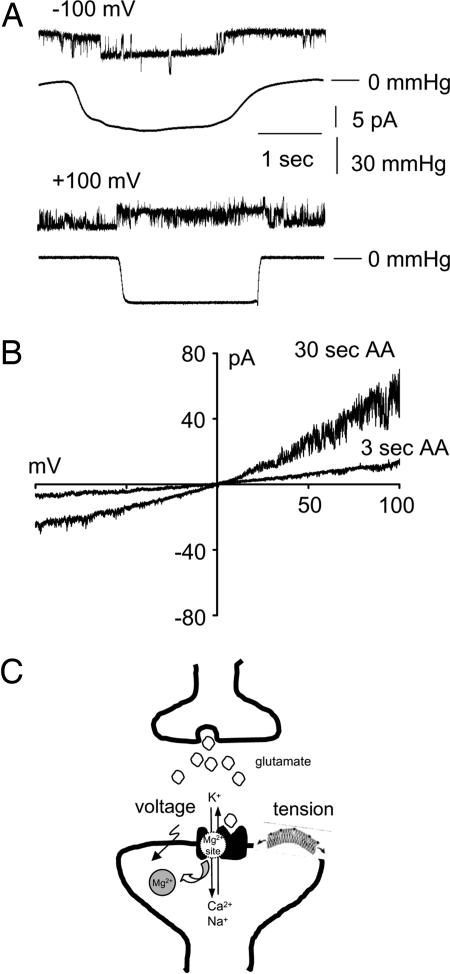
Fig. 4.
Modulation of Mg2+ block by stretch and arachidonic acid. (A) Stretch response of NMDA receptor channels. Representative currents were recorded from a liposome patch containing reconstituted recombinant NR1a and NR2A receptor subunits. The external pipette solution contained 10 μM glutamate, 100 μM glycine, 200 mM KCl, and 5 mM Hepes (pH 7.3). The internal bath solution facing the inside-out membrane contained 200 mM KCl, 5 mM MgCl2, and 5 mM Hepes (pH 7.3). The traces located below each current recording show the corresponding pressure recorded during applied suction (n = 5 patches). (B) Potentiation of NMDA receptor currents by arachidonic acid in isolated liposome membrane patches. Representative I–V curves for NR1a + NR2A subunit combinations obtained in the presence of 5 μM arachidonic acid evoked by voltage ramps from −100 mV to +100 mV. The recordings were acquired at 3 and 30 sec after the initial application of arachidonic acid to the liposome patch. The external pipette solution contained 10 μM glutamate, 100 μM glycine, 5 μM arachidonic acid, 200 mM KCl, and 5 mM Hepes (pH 7.3). The internal bath solution facing the inside-out membrane contained 200 mM KCl, 5 mM MgCl2, and 5 mM Hepes (pH 7.3) (n = 5 patches). (C) A schematic diagram of polymodal modulation of NMDA receptor currents. At the glutamatergic synapse, coincidental release of glutamate and membrane depolarization and/or membrane stretch releases the resting Mg2+ block, leading to ion influx.

