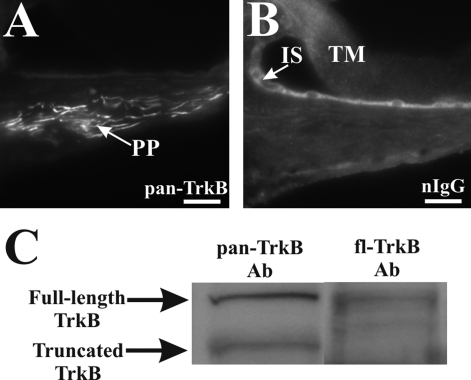
Figure 2-6921.
Characterization of the pan-TrkB antibody. Peripheral processes from a normal hearing rat cochlea showed a prominent expression of TrkB using the pan-TrkB (sc-8316) antibody (A), and no expression could be detected in these fibers located within the osseous spiral lamina area when a naïve immunoglobulin was used (B). In contrast, nonspecific binding of this antibody was observed in both the tectorial membrane (B, TM) and cells lining the inner sulcus (B, IS). Immunoblotting of cochlear proteins with the pan-TrkB antibody revealed the presence of both the full-length and truncated isoforms at ∼145 and ∼95 kd, respectively (C, left lane, pan-TrkB Ab), whereas immunoblotting with fl-TrkB antibody showed only the presence of the full-length isoform (C, right lane, fl-TrkB Ab). Scale bars= 20 μm. Original magnification, ×40.

