Figure 5-6921.
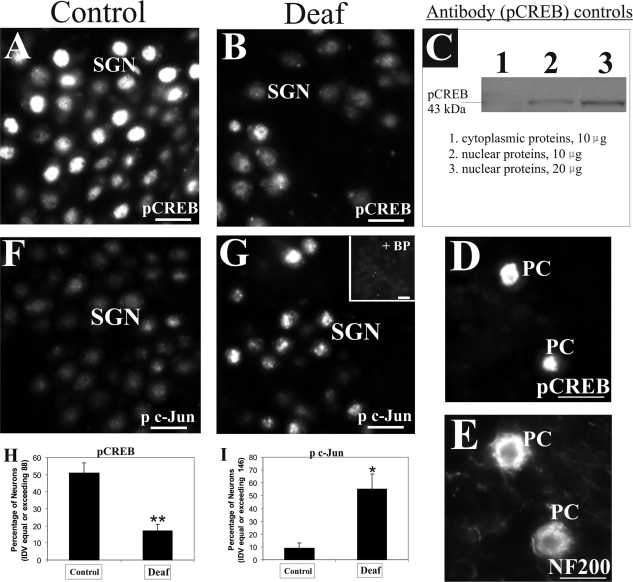
Aminoglycoside-induced sensorineural hearing loss induces differential phosphorylation of nuclear transcription factor in surviving SGNs. A: A large proportion of SGNs in normal hearing animals (left panel) expressed strong nuclear localization of pCREB protein in immunofluorescence studies. B: In deafened animals (middle panel), fewer neurons expressed such strong staining pattern of pCREB protein in their nuclei (as shown after 9 weeks of deafness). C: Characterization of pCREB antibody (right panel) revealed a single band of 43 kd in Western blots of nuclear proteins from hippocampal tissues (lane 2) but not in an equivalent amount of cytoplasmic proteins (lane 1). Furthermore, doubling the amount of hippocampal nuclear proteins led to a stronger band (compare lanes 2 and 3). To underline further the specificity of the antibody, immunolabeling of pCREB was observed in the nuclei of Purkinje neurons (D, PC) costained with NF200 to demarcate cytoplasmic regions (E, PC). F: In normal hearing animals, SGNs express low basal levels of p c-Jun protein in immunofluorescence analysis. G: However, deafened animals displayed elevation of p c-Jun fluorescence in their nuclei (shown here after 9 weeks of deafness), and most of this staining pattern was abolished by neutralization with the relevant control antigen (inset). Densitometric assessment of pCREB and p c-Jun fluorescence in the SGNs of four animals experiencing a deafness duration from 6 weeks to 3 months revealed a significant decrease in the proportion of SGNs demonstrating strong pCREB fluorescence (H, **P < 0.01), whereas in contrast, an increasing proportion of SGNs from these deafened animals demonstrated strong p c-Jun fluorescence (I, *P < 0.05). Numerical data indicate means ± SEM. Cochlear sections shown here were taken from the middle turn. Scale bars = 20 μm. Original magnification, ×40.

